Lamellar Ichthyosis
Autosomal recessive condition characterized by dark, plate-like adherent scales covering most of the body, typically presenting with a collodion membrane at birth.
What Is Lamellar Ichthyosis?
Autosomal recessive congenital ichthyosis (ARCI) is a recently adopted term referring to a heterogeneous group of disorders that share an autosomal recessive pattern of inheritance, collodion membrane presentation at birth and overlap in causative gene mutations.1 After shedding of the collodion membrane, skin of individuals with ARCI can show a variety of appearances. These different phenotypes have been termed lamellar ichthyosis (LI), congenital ichthyosiform erythroderma (CIE), and harlequin ichthyosis (HI). While significant overlap can occur between phenotypes and individual patients may manifest different phenotypes across their lifetime, these descriptors remain useful in classifying individuals with ARCI and often in predicting the underlying gene mutation.
Longterm Course
Lifelong; skin appearance may evolve early in life but generally stable thereafter; increased susceptibility to bacterial and fungal infections of skin; heat intolerance may be a problem for some.
Key Findings
- Skin
Generalized flat, polygonal, dark, often large (>1cm) scales; palms and soles usually thickened; variable redness under scales; ectropion usually present; may have clinical overlap with ARCI-CIE type due to shared gene mutations. - Hair
No shaft abnormalities; alopecia has been reported in some cases. - Nail
Usually normal; may become dystrophic.
Associated Findings
- Usually none
Clinical Resources
- Clinicians seeking to confirm a diagnosis should visit the Edvyce portal to submit a case to experts in ichthyosis.
- Learn more about FIRST’s Resources for individuals and families affected by Ichthyosis
- Information about current clinical trials and research studies can be found here.
What are the Signs & Symptoms?
ARCI is a rare disorder, estimated to occur in approximately 1 in 200,000 births.2 At birth, most individuals have a “collodion” presentation, so called because a clear membrane (the collodion) covers their bodies. Sometimes described as having a shellacked appearance, these newborns have skin that is taut, dark and split. Often the eyelids (ectropion) and lips (eclabium) can be forced open by the tightness of the skin, and there may be contractures around the fingers. The collodion membrane is gradually shed over the first few days to weeks of life, after which the skin can take a number of appearances. Such cases usually go on to develop either the LI or CIE phenotypes, with HI being comparatively rare.
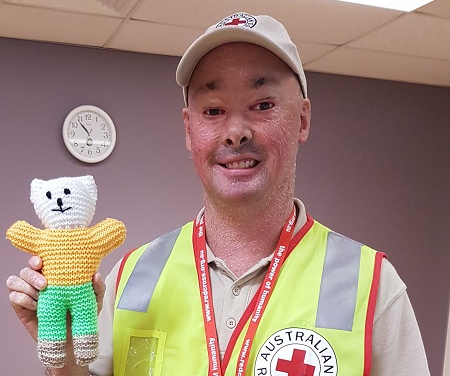
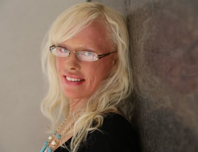
In individuals with the lamellar ichthyosis phenotype, shedding of the collodion membrane reveals dark, plate-like adherent scales with intervening cracks that cover most of the body surface. This build-up of scale is due to the fact that skin cells do not separate normally at the surface of the stratum corneum (the outermost layer of the skin) and are not shed as quickly as they should be. Even after shedding of the collodion membrane, people with lamellar ichthyosis often have trouble closing their eyes completely because of the tightness of the skin around the eyes and eyelids. In some cases, the skin around the eyes pulls so tightly it causes the eyelids to turn outward exposing the inner red lid and causing continuous irritation. This condition is called ectropion. If left untreated, damage to the cornea can develop leading to impaired vision. Treatment with topical retinoids can improve ectropion significantly, and surgical correction is now considered a treatment of last resort. People with lamellar ichthyosis may also have thickened nails and thickened skin on the palms and soles of their feet. Thick scale on the scalp may result in hair loss. Reddened skin underlying scale (erythroderma) is variable and reflects overlap with the ARCI-CIE phenotype. People with lamellar ichthyosis are at risk for superficial infections of their skin by fungus or bacteria as well as heat intolerance due to decreased sweating.
In individuals with the lamellar ichthyosis phenotype, shedding of the collodion membrane reveals dark, plate-like adherent scales with intervening cracks that cover most of the body surface. This build-up of scale is due to the fact that skin cells do not separate normally at the surface of the stratum corneum (the outermost layer of the skin) and are not shed as quickly as they should be. Even after shedding of the collodion membrane, people with lamellar ichthyosis often have trouble closing their eyes completely because of the tightness of the skin around the eyes and eyelids. In some cases, the skin around the eyes pulls so tightly it causes the eyelids to turn outward exposing the inner red lid and causing continuous irritation. This condition is called ectropion. If left untreated, damage to the cornea can develop leading to impaired vision. Treatment with topical retinoids can improve ectropion significantly, and surgical correction is now considered a treatment of last resort. People with lamellar ichthyosis may also have thickened nails and thickened skin on the palms and soles of their feet. Thick scale on the scalp may result in hair loss. Reddened skin underlying scale (erythroderma) is variable and reflects overlap with the ARCI-CIE phenotype. People with lamellar ichthyosis are at risk for superficial infections of their skin by fungus or bacteria as well as heat intolerance due to decreased sweating.
How is it Diagnosed?
The most common mutation in lamellar ichthyosis occurs in transglutaminase 1 (TGM1), an important enzyme for allowing skin to mature and eventually shed. Other causative mutations have been reported in ATP-binding cassette sub-family A member 12 (ABCA12), cytochrome P450 4F22 (CYP4F22), and ichthyin (NIPAL4).3 In other cases the gene mutation is not known. Mutations in lamellar ichthyosis usually (but not always) are transmitted through autosomal recessive inheritance. Individuals must inherit two recessive genes for lamellar ichthyosis to show the disease, one from each parent. Each parent (“carrier”) shows no evidence of lamellar ichthyosis. (For more information on the genetics of lamellar ichthyosis, request FIRST’s publication, Ichthyosis: The Genetics of its Inheritance).
Results of genetic tests, even when they identify a specific mutation, can rarely tell you how mild or how severe a condition will be in any particular individual. There may be a general presentation in a family or consistent findings for a particular diagnosis, but it’s important to know that every individual is different. The result of a genetic test may be “negative,” meaning no mutation was identified. This may help the doctor exclude certain diagnoses, although sometimes it can be unsatisfying to the patient. “Inconclusive” results occur occasionally, and this reflects the limitation in our knowledge and techniques for doing the test. But we can be optimistic about understanding more in the future, as science moves quickly and new discoveries are being made all the time. You can participate in research studies and also receive genetic testing through the National Ichthyosis Registry at Yale University or for more information about genetic tests performed you can visit GeneDx, www.genedx.com.
Results of genetic tests, even when they identify a specific mutation, can rarely tell you how mild or how severe a condition will be in any particular individual. There may be a general presentation in a family or consistent findings for a particular diagnosis, but it’s important to know that every individual is different. The result of a genetic test may be “negative,” meaning no mutation was identified. This may help the doctor exclude certain diagnoses, although sometimes it can be unsatisfying to the patient. “Inconclusive” results occur occasionally, and this reflects the limitation in our knowledge and techniques for doing the test. But we can be optimistic about understanding more in the future, as science moves quickly and new discoveries are being made all the time. You can participate in research studies and also receive genetic testing through the National Ichthyosis Registry at Yale University or for more information about genetic tests performed you can visit GeneDx, www.genedx.com.
What is the Treatment?
Lamellar ichthyosis is treated topically with skin barrier repair formulas containing ceramides or cholesterol, moisturizers with petrolatum or lanolin, and mild keratolytics (products containing alpha-hydroxy acids or urea). (For more information on which products contain these ingredients, refer to FIRST’s skin care products list.) For people with ectropion, in addition to treatment with topical retinoids, artificial tears or prescription ophthalmic ointments to lubricate the cornea are helpful in preventing eye damage. Severe lamellar ichthyosis can be treated systemically with oral synthetic retinoids (acitretin or isotretinoin). Retinoids are only used in severe cases of ichthyosis due to potential for effects on bones, tendons, and ligaments, and other complications.4,5
References
1. Oji V, Tadini G, Akiyama M et al. Revised nomenclature and classification of inherited ichthyoses: Results of the First Ichthyosis Consensus Conference in Sore`ze 2009. Journal of the American Academy of Dermatology. 2010; 63(4): 607-641.
2. Bale SJ, Richard G. Autosomal Recessive Congenital Ichthyosis. 2001 Jan 10 [Updated 2009 Nov 19]. In: Pagon RA, Bird TD, Dolan CR, et al., editors. GeneReviews™ [Internet]. Seattle (WA): University of Washington, Seattle; 1993-.
3. Fischer J. Autosomal Recessive Congenital Ichthyosis. J. Invest. Derm. 2009; 129:1319-1321.
4. Milstone L., McGuire J., Ablow R. Premature epiphyseal closure in a child receiving oral 13-cis-retinoic acid. J Am Acad Dermatol. 1982; 7:663-666.
5. Pittsley R.A., Yoder F.W. Retinoid hyperostosis: skeletal toxicity associated with long-term administration of 13-cis-retinoic acid for refractory ichthyosis. N Engl J Med. 1983; 308:1012-1014.
Types of Ichthyosis
- Congenital Ichthyosiform Erythroderma (CIE)
- Lamellar Ichthyosis
- Collodion Baby
- Darier Disease
- Epidermal Nevus
- Epidermolytic Ichthyosis
- Erythrokeratodermia Variabilis
- Erythrokeratodermia Cardiomyopathy Syndrome
- Harlequin Ichthyosis
- Ichthyosis Vulgaris
- Ichthyosis with Confetti
- Keratitis-Ichthyosis-Deafness (KID)
- Netherton Syndrome
- Pachyonychia Congenita
- Palmoplantar Keratodermas
- Sjögren-Larsson Syndrome
- X-Linked Ichthyosis
- Chanarin Dorfman Syndrome
- CHILD Syndrome
- Conradi-Hünermann
- Ichthyosis Hystrix with Deafness (HID) Syndrome
- Ichthyosis Hystrix Curth-Macklin
- Refsum Disease
- Trichothiodystrophy
Phenotype Names
Lamellar ichthyosis
EDD Name
TGM1-nEDD (~32–38% of ARCI)
NIPAL4-nEDD (~16%)
ALOX12B-nEDD (~12%)
CYP4F22-nEDD (~8%)
ABCA12-nEDD (~5%)
ALOXE3-nEDD (~5%)
PNPLA1-nEDD
CERS3-nEDD
SDR9C7-nEDD
SULT2B1-nEDD
LIPN-nEDD
CASP14-nEDD
OMIM
Inheritance
Autosomal recessive in most cases
Incidence
1:200,000
Diagnostic Tests
Genetic testing of blood
Age of First Appearance
Birth, usually
Abnormal Gene
transglutaminase 1 TGM1 in many cases; mutations also reported in ATP-binding cassette sub-family A member 12 ABCA12, cytochrome P450 4F22 CYP4F22, and ichthyin NIPAL4.
Primary Symptom Photos