
Molecular Mechanisms Linking Mechanical Stress to Epidermolytic Ichthyosis Severity (2025)

Xiaomin Bao, PhD
Associate Professor Northwestern University
Award: $50,000
For adult patients with epidermolytic ichthyosis, the most debilitating symptoms are localized to areas undergoing frequent mechanical stress, such as jonits and skin folds. We will investigate the molecular mechanisms underlying these localized symptoms, which will serve as a basis for treatment development.
AIMS:
The goal of this study is to determine how the skin cells from patients with epidermolytic ichthyosis (EI) respond to mechanical stress differently from healthy donors. We have developed two aims to determine changes in 1) the epigenome that predispose EI to heightened responses to mechanical stress; 2) cellular signaling pathways that respond to cyclic mechanical stretches.
METHODS:
We will leverage patient-derived primary human skin cells as the powerhouse to uncover the molecular mechanisms underlying EI’s augmented cellular responses to mechanical stress. Site-and age-matched skin cells from healthy donors will be used as controls.
RELEVANCE TO THE MISSION OF FIRST:
This study investigates the molecular mechanisms underlying the severe symptoms of EI that are specifically localized to joints and skin fold areas. The mechanistic insights generated from this study can inform the development of new targeted therapies. In particular, several FDA-approved drugs targeting components of the mechano-transduction pathway or epigenetic regulators are already available. Thus, the findings from this study will inform the possibilities of repurposing these drugs for EI treatment.
Beyond EI, findings from this study may provide a broader framework for understanding other subtypes of ichthyosis and related skin types with localized severity.
Progress Report: November 2026
Elucidating the Mechanistic Link between Epidermolytic Ichthyosis Severity and Mechanical Stress
Overview: Leveraging patient-derived primary human keratinocytes and my expertise in characterizing the molecular mechanisms in gene regulation, this research project is designed to uncover new mechanisms that can be targeted for treating epidermolytic ichthyosis (EI). We hypothesize that EI keratinocytes exhibit heightened responses to mechanical stress due to a combination of (1) epigenomic reprogramming resulting from years of adaptation to chronic mechanical stress and (2) enhanced cellular signaling in response to mechanical stimuli.
1. Expansion of primary keratinocytes from EI patients vs healthy donors
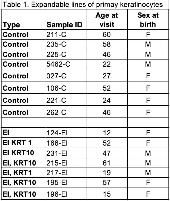
Primary patient-derived human keratinocytes are the foundation for this project. EI is a rare genetic disease. With the support from FIRST and the SBDRC (Skin Biology & Diseases Resource-Based Center) here at Northwestern University, we were able to successfully obtain and expand additional lines of primary cells in my laboratory for molecular mechanism characterization. To date, we have confirmed the expandability of primary keratinocytes isolated from seven EI patients, ranging in age from 12 to 61 years (3 males, 4 females). We also have confirmed expandability of control keratinocytes isolated from eight healthy donors, with age ranges from 24-60 (3 males, 5 females). All the keratinocytes were originally isolated from the same body location (upper inner arm). These cells are currently being used for different types of next-generation sequencing to profile their epigenetic landscape (details included in point #2) and response to mechanical cues (details included in point #3). The list of the expandable lines of primary patient-derived keratinocytes is included as Table 1
2. Elucidating the epigenetic components that contribute to EI disease severity.
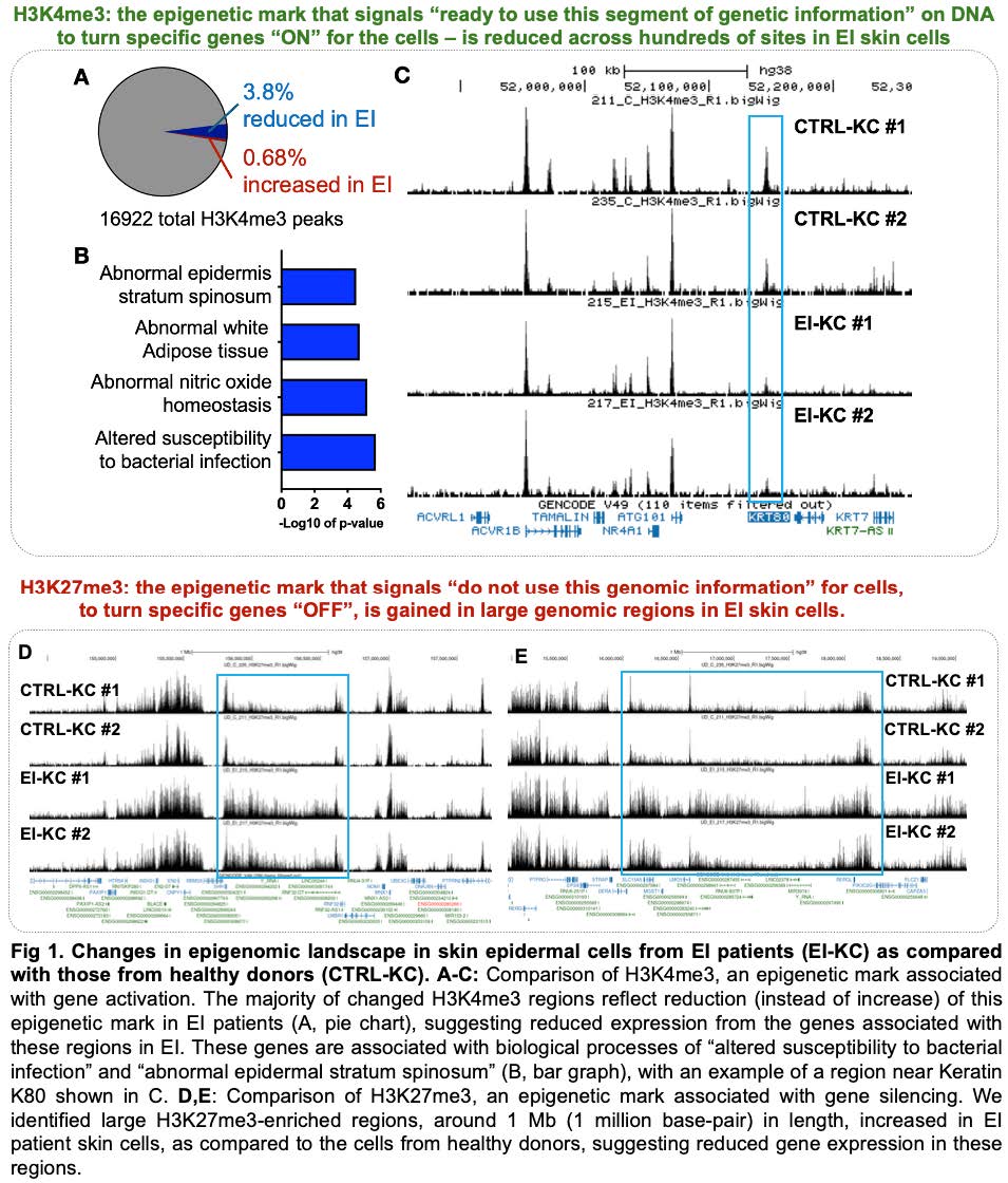
Considering the drastic changes in EI’s clinical manifestations from birth to later in life, we hypothesize that changes in the epigenetic landscape contribute to the adaptation of skin epidermis to EI fragility. While our initial findings were based on H3K4me3 and H3K27me3, from two controls and two EI patients, we are currently expanding our efforts to the following:
2.1. Including Additional epigenetic markers and RNA-seq
Building on the consistent changes we observed in H3K4me3 (epigenetic mark for active promoters) and H3K27me3 (epigenetic gene silencing controlled by the Polycomb complexes, often reversible), (details included in Fig.1), we are currently including H3K27Ac (epigenetic mark for active enhancers), H3K9me3 (stable gene silencing and genome protection), H2AZ (epigenetic memory) in our epigenomic profiling endeavors. Because the published RNA-seq data is based on entire skin biopsies, which contains a mixture of cell types and cell states, we have been also generating RNA- seq data for keratinocytes at the matched cell states with the epigenomic data sets. These efforts are designed to provide a comprehensive overview of altered epigenomic landscape in EI patients as compared to normal healthy donors, and to connect the changes in epigenetics to state-specific gene expression in keratinocytes.
2.2 Testing alternative epigenomic profiling approaches
Our initial approach for profiling epigenomic marks was Cut&Run, which requires 0.5 million per replicate. While we are able to expand primary keratinocytes in vitro, their capacity to proliferate is still very limited. To accommodate the increased number of epigenomic markers that we want to profile, we are currently investigating Cut&Tag as an alternative approach. Cut&Tag only requires 0.1 million cells per replicate, and can greatly increase our throughput once we fully optimize the conditions in keratinocytes.
3. Investigating the differential mechanical sensitivity in keratinocytes between EI and healthy donors
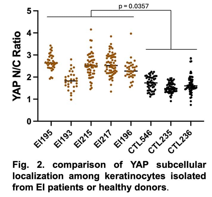
The severity of EI in joint areas suggest that the keratinocytes exhibit heightened response to mechanical stress. In parallel with our development of a custom-built stretching device (reported in April), we have established an orthogonal approach using substrates of varying stiffness. PDMS cured at the ratio of 1:15 has an estimated stiffness of 0.8-1.0 MPa Young’s modulus, mimicking the stiffness of human skin. We use the subcellular localization of YAP1, a key mechano-transducing transcription co-activator that senses mechanical cues and drives cellular proliferation, as a readout for keratinocytes’ mechanosensation.
As shown in Fig. 2, we have quantified YAP1 subcellular localization across five lines of EI and 3 lines of control keratinocytes. Keratinocytes isolated from EI patients consistently exhibit higher nuclear to cytoplasmic ratio of YAP, as compared to keratinocytes isolated from healthy donors. This increased nuclear localization (p=0.0357, Exact permutation test) suggests heightened YAP activity that can promote keratinocyte proliferation. We are currently testing if YAP inhibition can reduce proliferation especially in the keratinocytes isolated from EI patients.
4. Additional progress.
We have presented our preliminary findings in the FIRST 2026 conference as a poster. We have also submitted an R21 to NIH and received the rating of “competitive but not discussed”. We plan to gather additional data, publish the first paper, and submit an R01. In terms of personnel, we have successfully recruited an outstanding new graduate student in the PhD program to drive this project. We are grateful for the funding from FIRST, and we are confident that our team will be able to make meaningful contributions to improve the quality of life for the patients with ichthyosis.

If you are a physician who treats patients with ichthyosis and you would like to be added to our listing, complete this form.