Epidermal Nevus: A Patient's Perspective: A Clinical Perspective
What is Epidermal Nevus?
Epidermal nevus is a clinical term for a family of skin lesions that involve the outer portion of skin, the epidermis, and are distributed in a linear and often swirled pattern. Overall, epidermal nevi are not uncommon congenital malformations, occurring in 1-3 per 1000 births.
What are the Signs & Symptoms?
The lesions may be single or multiple and are usually present at birth. All epidermal nevi show some changes in texture which can range from very rough, warty and spiny, and often darker than the surrounding normal or uninvolved skin (verrucous epidermal nevus), to red and scaly (inflammatory linear verrucous epidermal nevus or ILVEN), to yellowish, rough and pebbly appearance due to proliferation of oil- or ’sebaceous’ gland-like structures (nevus sebaceous). If they are limited to the epidermal linings of the hair follicles, they may appear like blackheads: nevus comedonicus. Another related and relatively common entity, the Becker’s nevus, is a form of epidermal nevus with a somewhat velvety texture, mild to moderate hyperpigmentation and coarser vellus hairs. It typically occurs on the trunk during childhood or adolescence in a broader, patchy rather than linear pattern. The term ‘nevus’ is also used for other birthmarks, malformations and some benign growths, such as melanocytic nevi, or ‘moles’.
How is it Diagnosed?
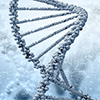
Epidermal nevi are genetically ‘mosaic’, meaning that the mutation causing the nevi are not found in other cells of the body. Mosaicism arises when the genetic mutation occurs in one of the cells of the early embryo sometime after conception; such mutations are called ‘somatic’ mutations. This mutated cell, like the other normal cells, continues to divide and gives rise to mutated daughter cells that will populate a part of the body. The linear patterning of the epidermal nevus reflects the movement of the mutant daughter cells during fetal growth. These linear, developmental patterns are termed the ‘lines of Blaschko’. Many epidermal cells within these affected areas harbor the mutant gene, while most or all cells from uninvolved areas do not. After birth, the nevus “grows with the child”, although some new areas of involvement and/or extension of the nevus to new areas can occur.
ILVEN is an exception: here, lesions often do not appear until later in infancy or childhood. Some other genetic conditions that have linear, swirled patterning, similar to an epidermal nevus, do not arise because of genetic mosaicism. These include CHILD syndrome, Conradi-Hünermann-Happle syndrome, and incontinentia pigmenti. Instead, these disorders represent “functional mosaicism”, where the mutant gene is in every cell, but that gene is active in only parts of the body. The patterned distribution reflects gene activity (i.e., DNA transcribed into RNA which is translated into protein) in the affected areas, and inactivity in the unaffected areas.
Epidermal nevus syndrome and its subtypes, nevus sebaceous syndrome (syn: Schimmelpennig-Feuerstein-Mims syndrome) and nevus comedonicus syndrome, occur when some epidermal nevi are associated with defects or malformations in other organ systems, particularly of the central nervous system, eyes, and the skeleton. Rarely, patients may also present with hypophosphatemic rickets (cutaneous skeletal hypophosphatemia syndrome).
Another variant of epidermal nevus syndrome is phakomatosis pigmentokeratotica, in which both epidermal nevi and congenital pigmented lesions are present on different parts of the skin surface. Yet another variant, Becker’s nevus syndrome, presents with breast and/or skeletal hypoplasia in the same region as the nevus.
Several other genetic disorders display linear or segmental keratotic lesions, and are sometimes grouped with epidermal nevi. Proteus syndrome is a multisystem disorder characterized by overgrowth of multiple tissues. Features are highly variable between patients, and include nevi of epidermal and connective tissue origin, hemihypertrophy, lipomas, vascular malformation, asymmetrical macrodactyly, and tumors of the ovaries or parotids. Linear forms of Darier disease are reported, which present with a stripe of classic Darier’s lesions, with occasional nail abnormalities on one side of the body. The lesions have histopathology consistent with Darier’s and are found to have somatic mutations in ATP2A2,the causative gene for Darier disease. Porokeratotic eccrine nevus is another keratotic epidermal nevus. It involves the intraepidermal ducts of the eccrine (sweat) glands with a characteristic histopathology. Lesions may be limited to palms/soles or more widespread in a linear pattern and are due to somatic mutations in GJB2.
Some linear keratotic disorders like Porokeratosis of Mibelli represent another form of genetic mosaicism: type 2 mosaicism, or type 2 segmental manifestation.
This occurs in autosomal dominant disorders, where heterozygous individuals (having 1 mutant copy and 1 normal copy of a gene) have diffuse disease throughout the body, superimposed with segments or lines of more severe lesions. These segments are mosaic areas where the normal copy of the gene was lost or deleted, known as “loss of heterozygosity”. Without a normal copy of the gene to buffer the mutant copy, the disease becomes more severe in these regions.
The genetics of epidermal nevi, including the sebaceous and comedonal subtypes, reflect this clinical heterogeneity. To date, mutations in keratin 1 (KRT1), keratin 10 (KRT10), fibroblast growth factor receptor 3 (FGFR3), fibroblast growth factor receptor 2 (FGFR2), PICK3A, HRAS, KRAS, and NRAS have been identified. Genetic diagnosis is performed on a skin biopsy from an affected area. For widespread or syndromic nevi, the same mutation is found in both the epidermal nevus and the other affected tissues, including other types of skin lesions. It is likely that in the future with more research, it will be possible to associate particular patterns of malformations with specific genes. Parents of a child with an epidermal nevus should generally not be at increased risk for other affected children, because the mutation did not arise within their sperm or eggs, but within the fetal tissue. However, there is some risk that the individual with an epidermal nevus could pass the mutant gene onto their offspring, if their germline tissue is also mosaic for the mutation. For example, affected offspring with widespread epidermolytic ichthyosis has been reported from individuals with epidermolytic epidermal nevi. This risk is difficult to quantify, and patients desiring more information are advised to seek formal genetic counseling. Because of the high probability that these nevi carry a mutation in either keratin 1 or keratin 10, skin biopsy to look for features epidermolytic hyperkeratosis may also be useful prior to genetic testing.
Linear or “segmental Darier disease” similarly carries risk for offspring with generalized or complete Darier disease (due to mutations in ATP2A2;. Somatic mutations in GJB2, the gene encoding connexin 26, are reported in porokeratotic eccrine nevus. Mutations in this same gene cause keratitis-ichthyosis-deafness (KID) syndrome, suggesting that patients with this nevus might carry some risk for offspring with KID syndrome. The autosomal dominant Porokeratosis of Mibelli was attributed to a mutation in mevolonate kinase (MVK) in one kindred. Mutations in PTEN and AKT1 underlie Proteus syndrome. The genetic bases for ILVEN and Becker’s nevus are yet unknown.
Doctors frequently use genetic testing to help define which ichthyosis a person actually has. This may help them to treat and manage the patient. Another reason to have a genetic test is if you or a family member wants to have children. Genetic testing, which would ideally be performed first on the person with ichthyosis, is often helpful in determining a person's, and their relative's, chances to have a baby with ichthyosis. Genetic testing may be recommended if the inheritance pattern is unclear or if you or a family member is interested in reproductive options such as genetic diagnosis before implantation or prenatal diagnosis.
Results of genetic tests, even when they identify a specific mutation, can rarely tell how mild or how severe a condition will be in any particular individual. There may be a general presentation in a family or consistent findings for a particular diagnosis, but it's important to know that every individual is different. The result of a genetic test may be "negative," meaning no mutation was identified. This may help the doctor exclude certain diagnoses, although sometimes it can be unsatisfying to the patient. "Inconclusive" results occur occasionally, and this reflects the limitation in our knowledge and techniques for doing the test. But we can be optimistic about understanding more in the future, as science moves quickly and new discoveries are being made all the time. You can participate in research studies and also receive genetic testing through the National Ichthyosis Registry at Yale University or for more information about genetic tests performed you can visit GeneDx, www.genedx.com.
Download a PDF version of this information
References.
1. Sugarman JL. Epidermal Nevus Syndrome. Semin Cutan Med Surg 2007;26:221-230.
2. Paller AS, Syder AJ, Chan YM et al. Genetic and clinical mosaicism in a type of epidermal nevus. N Engl J Med 1994;331:1408-1415.
3. Tsubota A, Akiyama M, Sakai K et al. Keratin 1 gene mutation detected in epidermal nevus with epidermolytic hyperkeratosis. J Invest Dermatol 2007;127:1371-1374.
4. Lim YH, Ovejero D, Sugarman JS et al. Multilineage somatic activating mutations in HRAS and NRAS cause mosaic cutaneous and skeletal lesions, elevated FGF23 and hypophosphatemia. Hum Mol Genet 2014;23:397-407.
5. Li JY, Berger MF, Marghoob A et al. Combined melanocytic and sweat gland neoplasm: cell subsets harbor an identical HRAS mutation in phacomatosis pigmentokeratotica. J Cutan Pathol 2014;41:663-671.
6. Harboe TL, Willems P, Jespersgaard C et al. Mosaicism in segmental Darier disease: an in-depth molecular analysis quantifying the proportions of mutated alleles in varous tissues. Dermatol 2011;222:292-296.
7. Zeng K, Zhang QC, Li L et al. Splicing mutation in MVK is a cause of Porokeratosis of Mibelli. Arch Dermatol Res 20ar;306:749-755.
8. Paller AS. Piecing together the puzzle of cutaneous mosaicism. J Clin Invest. 2004;114:1407-1409.
9. Zhou X_P, Marsh DJ, Hampel H et al. Germline and germline mosaic PTEN mutations associated with a Proteus-like syndrome of hemihypertrophy, lower limb asymmetry, arteriovenous malformations and lipomatosis. Hum Mol Gen 2000;9:765-768.
10. Lindhurst MJ, Sapp JC, Teer JK, et al. A mosaic activation mutation in AKT1 associated with the Proteus Syndrome. N Engl J Med 2011;365:611-619.
| Other Names: | nevus sebaceous, verrucous epidermal nevus, keratinocytic epidermal nevus, inflammatory linear verrucous epidermal nevus (ILVEN), Becker’s nevus, Porkeratotic Eccrine and Ostial Dermal Duct Nevus. |
| OMIM: | 162900 |
| Inheritance: | sporadic, somatic mutation |
| Incidence: | 1 to 3 per 1000 births |
| Key Findings: |
|
| Associated Findings: |
|
| Age at First Appearance: | Usually birth. Typically delayed to childhood or adolescence in ILVEN and Becker’s nevi. |
| Longterm Course: | The nevus usually “grows with the child”; new areas of involvement or extension of existing nevus to new areas can sometimes occur. |
| Diagnostic Tests: | genetic testing of affected tissues. |
| Abnormal Gene: | ATP2A2, KRT1, KRT10, FGF2, FGF3, GJB2, HRAS, NRAS, KRAS, PICK3A, |
Additional Resources:
<
- Clinicians seeking to confirm a diagnosis should visit FIRST's TeleIchthyosis site to submit a case to experts in ichthyosis. »
- Learn more about FIRST's Support Services - connecting affected individuals and families with each other. Or call the FIRST office at 800-545-3286. »
- Information about current clinical trials and research studies can be found here.